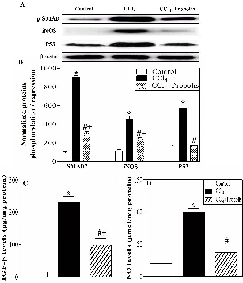
Fig. 7. Induction of liver fibrosis was associated with alterations in TGF-β, P53 and iNOS signaling. Liver lysates were prepared from hepatic tissues of mice from each group. The liver lysates were then subjected to Western blotting using antibodies recognizing pSMAD2, P53, iNOS and β-actin. The protein bands from one representative experiment are shown for the phosphorylation of SMAD2 and the expression of iNOS, P53 and β-actin (A). The phosphorylated SMAD2 and the expression of iNOS and P53 were normalized to the total β-actin protein levels. The results are expressed as the means ± SEM of the normalized value of p-SMAD2 in the three animal groups, (n=5) (B). The levels of TGF-β were measured in the liver lysates from control (open bar), CCl4-treated (closed back bar) and CCl4+propolis-treated (hatched bar) groups by ELISA. The results of five individual mice from each group are expressed as the mean level of TGF-β ± SEM (C). The levels of NO were also measured in the liver lysates of five mice from each group, and the results are expressed as the mean level of NO ± SEM (D).